
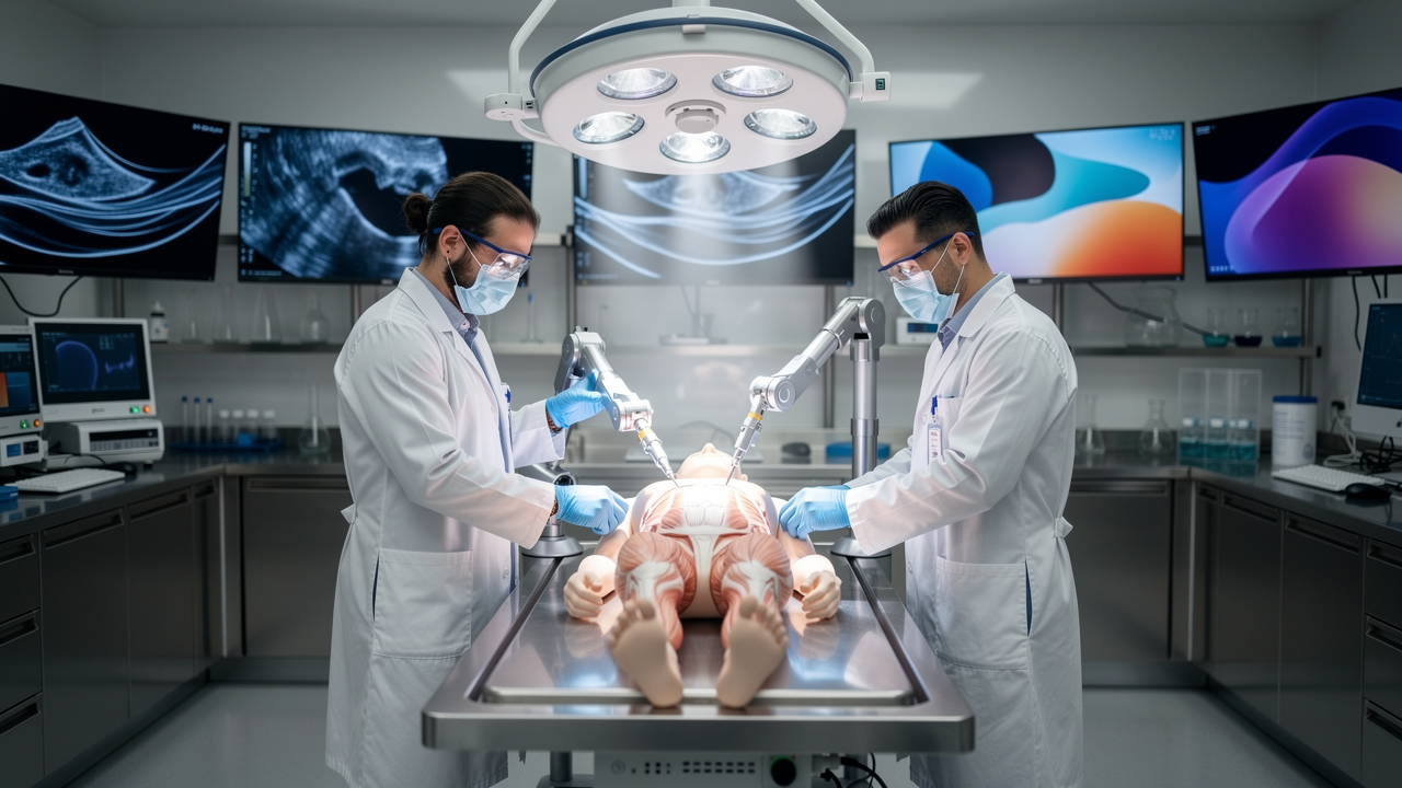
What is cadaver-based pre-clinical testing in medical device development?
Cadaver-based pre-clinical testing is the validation of surgical instruments, implants, and medical devices using human tissue specimens before those devices reach clinical trials or human patients. It sits at a specific point in the medical device development process, offering engineers a chance to interact with real anatomy rather than synthetic approximations. A human cadaver model allows a device team to observe how a product behaves inside tissue layers, joints, and vascular structures that no digital simulation fully replicates.
This method functions as a bridge between benchtop testing and the clinical environment. Nonclinical device testing conducted on cadavers exposes issues that would otherwise surface much later, during simulated use testing or even during live procedures. Ex vivo testing and human tissue testing are often grouped together, but cadaveric work specifically emphasizes anatomical accuracy that other ex vivo methods cannot match.
Where cadaveric testing fits in the development pathway
Proof of concept testing typically confirms that a device performs its basic function under controlled, artificial conditions. Cadaveric testing picks up after that stage, once mechanical validation is complete, and before in vivo animal testing or human clinical trials begin. This sequencing matters: a device that passes benchtop checks may still fail when navigating real anatomical variation, and catching that gap early avoids wasted downstream effort.
Which devices benefit most from ex vivo testing
Complex, anatomy-dependent systems gain the most from this approach. Orthopedic implants need exact bone contour matching. Endovascular catheters must navigate vessel tortuosity that differs from patient to patient. Robotic surgical consoles require realistic tissue resistance to calibrate force feedback. Surgical device evaluation on cadavers gives engineers a much clearer picture of how these systems behave against structures that synthetic models cannot fully replicate.
Why Cadaver-Based Pre-Clinical Testing Accelerates Medical Device Development
Using human cadavers speeds up research and development by letting engineering teams catch design flaws and usability problems early, before they become expensive late-stage redesign cycles. Physical feedback from human specimens is immediate, a poor grip angle, an instrument that catches on tissue, or a delivery system that binds inside a vessel becomes obvious within a single test session. This directness supports faster design iteration and shortens early feasibility testing timelines considerably.
Faster design iteration and prototype evaluation
In a cadaver lab testing environment, engineers can test several design modifications across a single weekend session. Compare that to animal model protocols, which often require weeks of scheduling, ethical approval, and recovery observation. Immediate prototype evaluation during mock surgeries means a design flaw identified on a Friday can be corrected and retested by Sunday.
Earlier identification of design failure points
Realistic mechanical stress and anatomical constraints reveal failure points in delivery systems and hand instruments that bench testing simply does not simulate. A catheter that kinks against a synthetic tube may behave differently against a genuinely tortuous vessel. This kind of risk reduction in development depends on anticipating clinical failure modes while the device is still in ex vivo tissue, not after it has entered a patient.
What can cadaveric testing reveal that traditional bench testing cannot?
Benchtop physical models and synthetic phantoms lack the natural variation, compliance, and multi-layered tissue resistance found in real human anatomy. A silicone vessel model might approximate diameter, but it rarely reproduces the friction, elasticity, and layered resistance of an actual artery wall. Tactile feedback during instrument insertion, the friction of tissue against a device surface, and hidden architectural constraints such as adjacent nerve bundles or fascial planes only appear in genuine anatomical testing.
A scientific comparison of preclinical models focused on mechanical thrombectomy devices found that cadaveric specimens revealed procedural performance issues tied to vessel tortuosity and anatomical variation that phantom and animal models did not fully capture. That kind of peer-reviewed research on device testing models supports the case for cadaveric evaluation as a distinct and necessary step rather than an optional add-on to bench work.
Assessing device-tissue interaction and anatomical realism
Vascular pathways, bone density variations, and fascial planes all behave differently under mechanical stress than their synthetic counterparts. Anatomical realism affects tactile forces experienced by the operator and the accuracy of simulated surgical navigation. A device that tracks smoothly through a straight synthetic tube may behave unpredictably in a vessel with natural curvature and variable wall thickness.
Simulating clinical workflow and surgical access
Beyond the device itself, cadaveric sessions expose physical constraints that only appear in a realistic simulated operating room: line-of-sight issues, instrument crowding around a surgical field, and awkward hand positioning. Clinical workflow simulation shows how clinicians interact with a device interface under the same physical pressures they would face during an actual procedure, including fatigue and limited access angles.
How does cadaver testing compare to animal testing for medical devices?
Animal models remain useful for evaluating biological responses, healing patterns, and systemic compatibility. Human specimens, on the other hand, offer structural realism that animal tissue cannot replicate, particularly for devices designed around human-specific pathologies. Animal anatomy frequently lacks matching structural geometry, which makes direct implant fit assessment difficult or misleading when relying solely on animal testing.
Pre-clinical medical device testing increasingly treats cadaveric and animal work as complementary rather than interchangeable. Reviewing the preclinical development phase on a general level helps clarify why regulatory frameworks often expect multiple testing modalities rather than a single validation method before a device advances toward clinical trials.
Evaluating implant fit assessment and human anatomy realism
Orthopedic implants and cardiovascular devices depend on human bone contours and vessel dimensions that vary meaningfully from animal analogues. Human tissue testing prevents design errors that stem from species-specific anatomical differences, such as a joint replacement sized around canine or porcine bone structure that does not translate accurately to human skeletal geometry.
Understanding when animal models remain necessary
Animal models still play a necessary role in assessing perfusion, biocompatibility, local inflammatory response, and long-term healing profiles, factors that cadaveric tissue cannot demonstrate since it lacks living biological processes. Combining both methodologies, ex vivo validation on human specimens paired with in vivo animal studies, forms a translational testing strategy that covers structural and biological questions together.
How does this method reduce costly redesigns and development delays?
Discovering a design mismatch during live clinical trials can set a project back by months or years and cost a device company a substantial sum in redesign, retesting, and delayed market entry. Finding that same mismatch under ex vivo conditions resolves safety and usability questions well before final manufacturing decisions are locked in, when changes are still comparatively inexpensive to make.
Enhancing product development acceleration through early feedback
Failing fast and cheaply in a controlled testing environment carries clear financial benefits. A flawed delivery mechanism identified in a cadaver lab costs a fraction of what the same discovery would cost after a clinical trial has already begun. Rapid modifications made at this stage compress overall development timelines rather than extending them.
Integrating usability feedback from physician experts
Qualitative feedback from key opinion leaders participating in surgeon training environment sessions often surfaces issues that engineering teams alone would miss, such as an instrument handle that feels awkward during extended procedures. Converting surgeon comments into updated design requirements at this stage helps prevent market adoption failures after launch, when redesign is far more disruptive.
All the information awaits you at the following link: biotechanatomy.co.il.
What should a high-quality cadaver testing program include to ensure regulatory readiness?
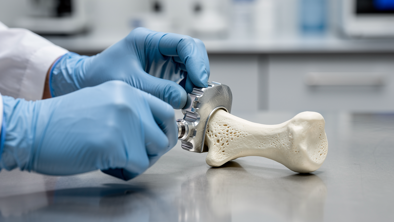
A successful testing run requires more than tissue procurement. It needs structured protocols, clear data collection tools, and traceable engineering parameters capable of satisfying regulatory bodies reviewing a submission. Proper documentation matters as much as the testing itself, since regulators expect a clear record connecting test observations to a device's risk file.
| Program Element | Purpose | Regulatory Relevance |
|---|---|---|
| Quantitative endpoints | Measure insertion force, placement accuracy | Converts observation into premarket evidence |
| Structured protocols | Standardize test conditions across sessions | Supports procedural validation |
| Physician feedback logs | Capture usability and workflow observations | Feeds design control updates |
| Traceability documentation | Link findings to design inputs | Builds verification planning history |
Protocol design and rigorous premarket evidence generation
Testing protocols built around quantitative endpoints, such as insertion forces or placement accuracy metrics, turn an experimental cadaver session into objective premarket evidence. Without measurable endpoints, a test session produces impressions rather than data that a regulatory reviewer can evaluate.
Aligning simulation studies with design controls and verification planning
Findings from ex vivo clinical workflow simulation need to map directly back to design inputs and risk management files. Documenting each design refinement establishes a development trail that shows regulators how a device evolved in response to real anatomical testing, rather than presenting a finished product with no traceable history.
Does cadaver testing replace the need for animal studies?
No. Cadaver testing addresses structural and anatomical fit, while animal studies address biological response, healing, and perfusion. Most development programs use both.
How many devices can be tested in one cadaver lab session?
This depends on the complexity of the device and the anatomical region involved, but multiple design iterations are often evaluated within a single session, which is far faster than sequential animal testing rounds.
Is cadaveric testing required by regulatory bodies?
Requirements vary by device classification and region, but many regulatory pathways expect some form of nonclinical device testing that includes anatomically realistic evaluation before clinical trials proceed.
What is the difference between ex vivo testing and in vivo testing?
Ex vivo testing, including cadaveric work, uses tissue outside a living biological system. In vivo testing occurs within a living organism, such as an animal model, and captures biological responses that ex vivo methods cannot.
About the Business
Biotech Anatomy LTD operates a state-of-the-art facility dedicated to practical anatomy and surgical education, supporting surgeons, medical students, healthcare professionals, researchers, and medical device development companies through cadaver-based training and research and development services. The facility provides advanced laboratory settings paired with radiological imaging integration and precise simulated operating environments, alongside high-quality human anatomical specimens suited to detailed pre-clinical work. Its programs facilitate physician feedback sessions, clinical workflow simulation, and structured pre-clinical testing protocols designed to reduce risk in medical technology projects and shorten development timelines for device teams working toward regulatory milestones.